MiraQ™ Cardiac
Improve surgical outcomes, demonstrate quality, and increase cost-efficiency.
Combining the spatial information from epicardial ultrasound imaging and quantitative data from TTFM enables the surgeon to perform a prompt and accurate assessment, and revise the graft when necessary.
The Medistim MiraQ™ Cardiac combines ultrasound imaging and transit time flow measurement (TTFM) in a single system that is specifically designed for cardiac surgery.
There is growing support of the idea that checking grafts and anastomoses during cardiac surgery should be standard of care. Reliability and ease of use is a major determinant for this to become reality.
The MiraQ™ Cardiac system has built-in support for Guided Workflows. These are software protocols that assist the user to a standardized approach to quality assessment. Intraoperative quality assessment has become easier to adopt, customizable to the surgeon’s needs and enhancing work efficiency.

Epicardial imaging
Epicardial ultrasound imaging gives a simple, fast and safe imaging of coronary stenoses and graft anastomoses, providing immediate feedback on the quality of the CABG surgery.
Transit Time Flow Measurement
Performing flow measurements with the MiraQ™ Cardiac is the quickest and most accurate way to verify graft patency while the patient is still in the operating room.
Epiaortic imaging
Epiaortic imaging provides a sensitive, direct diagnosis of aortic disease. This may lead to modifications of the surgical strategy and thus contribute towards reduced rates of major adverse cardiac and cerebrovascular events (MACCE) following surgery.
Combining imaging and flow for better quality assessment
The MiraQ™ Cardiac system uses Medistim’s flow measurement and high-resolution ultrasound Imaging probes to provide a complete quality assessment.
Medistim’s L15 High-frequency ultrasound Imaging probe provides high-resolution images that allows the surgeon to asses morphology. Medistim’s flow probes utilize transit time technology to accurately measure blood volume flow intraoperatively.
Combining the spatial information from epicardial ultrasound imaging and quantitative data from TTFM enables the surgeon to perform a prompt and accurate assessment, and revise the graft when necessary
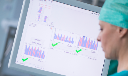
Instant feedback
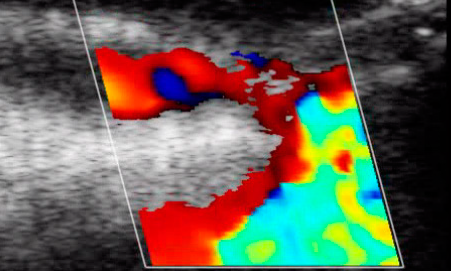
The MiraQ™ Cardiac provides instant feedback on the performance of a graft. Eliminate guesswork with ultrasound imaging visualization and quantifiable TTFM data.
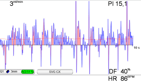
In the SVG-CX measurements presented here, ultrasound imaging was used to scan both the distal and proximal anastomosis for defects. An occluded proximal anastomosis was discovered and verified by a TTFM measurement (PI 15.1, DF 40% and Flow 3 ml/min
Revise on the spot
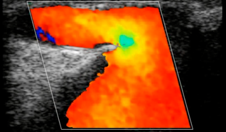
When occluded or underperforming grafts are detected they can be revised on the spot. Take every measure to avoid recalls.
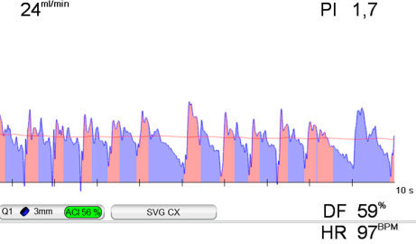
The occluded SVG-CX was immediately revised, and the improved result was documented with ultrasound imaging and TTFM. As shown above, the graft flow was significantly improved (PI 1.7, DF 59% and Flow 24 ml/min).
TTFM Reliable flow volume measurement
The established numeric indices Pulsatility Index (PI), Diastolic Filling (DF%) and Mean Flow, the basis of our 3-parameter assessment method, provide an accurate insight into the dynamics of graft function. TTFM is included in the guidelines endorsed by EACTS/ESC, NICE, and STS.