MiraQ™ Vascular
Improve surgical outcomes, demonstrate quality and increase cost efficiency.
Combining the spatial information from epicardial ultrasound imaging and quantitative data from TTFM enables the surgeon to perform a prompt and accurate assessment, and revise the graft when necessary.
MiraQ™ Vascular combines ultrasound imaging and transit time flow measurement (TTFM) in a single system that is specifically designed to meet the needs of vascular surgery.
Performing perioperative quality assessment using Medistim technology can greatly increase the patients’ probability of a positive outcome and lessen the chance of additional and unnecessary surgical reinterventions.
The MiraQ™ Vascular system provides objective, quantifiable feedback on how well a graft is functioning during an operation. Surgeons can leave the operating room with the assurance that the construct is functioning well. All surgical findings can be documented through the flow tracings and images provided by the Medistim system.

Carotid Endarterectomy (CEA)
The MiraQ™ Vascular system offers the unique combination of high frequency ultrasound imaging guidance and flow measurement (TTFM). Documented to reduce risk of death and stroke and improve cost effectiveness.
Peripheral bypass surgery
With graft patency being the predominant predictor of long-term graft survival, surgeons can improve patient quality of life and reduce reinterventions, using the MiraQ™ Vascular system.
AV Access
Flow quantification and intraoperative guidance are valuable tools for performing AV Access surgery. This improves the probability of a long lasting well-functioning shunt, reduce the risk of cardiac failure and hand ischemia.
Combining imaging and flow for better surgical guidance and quality assessment
Medistim’s L15 High-frequency ultrasound Imaging probe provides high-resolution images that allows the surgeon to asses morphology. Medistim’s flow probes utilize transit time technology to accurately measure blood volume flow intraoperatively.
Combining the spatial information from ultrasound imaging and quantitative data from TTFM enables the surgeon to make informed decisions, and revise grafts when necessary.
Intraoperative Guidance
The MiraQ™ Vascular provides a comprehensive overview of the situation at hand.
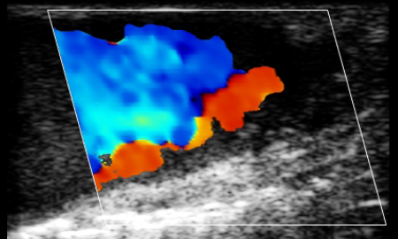
Ultrasound imaging is a valuable tool for visualization and evaluation of the stenosis and the completed endarterectomy
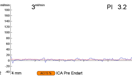
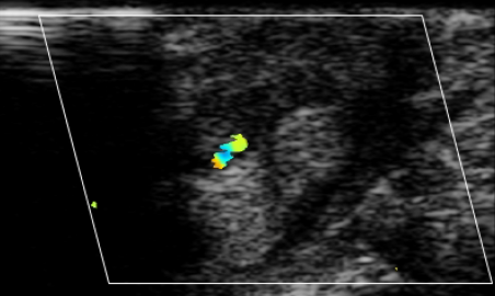
In the CEA procedure presented here, ultrasound imaging and TTFM was used to verify the location and severity of a stenosis prior to endarterectomy.
The color flow indicates little or no flow, and this is verified by a TTFM measurement. Note that the TTFM measurement has very low acoustic coupling index (ACI) due to the large amount of plaque in the vessel.
Verification While in the OR
An image of the carotid artery can reveal otherwise unseen imperfections and give the surgeon a chance to take appropriate actions.
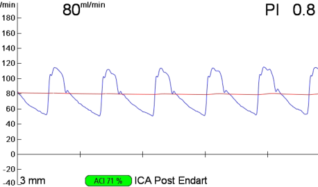
The above post-endarterectomy measurements clearly show a successful removal of the stenosis and a greatly improved flow.